Kidney Stone or Renal Colic or Urinary Stone Disease
Synonyms of Kidney Stones
- Renal Colic or Calculi
- Ureteric Colic
- Nephrolithiasis
- Urolithiasis
What are Kidney Stones or Renal Calculi?

They are basically deposits, which are made of minerals and salts, formed in the kidney.
They are hard and made of different minerals which form different types of stones.
It affects 240,000 to 720,000 Americans every year and while it affects men more than women in a ratio of 2.5:1, the incidence among females is slowly increasing. Patients are usually in their third to fifth decade but can happen at any age.
Prevalence of Kidney stones in the World
Prevalence in different parts of the world is as follows,
- USA : 13% to 15%,
- India : 9% to 10%.
- Europe : 5 to 9%
- South America: 3% to 5%.
- Australia : 7%
- North Asia : 1% to 8%
- South Asia : 5% to 9%
- East Asia : 1% to 8%
- West Asia : 5% to 9%
Types of Kidney Stones
- Calcium oxalate
- Calcium Phosphate
- Struvite ( Magnesium ammonium Phosphate)
- Uric Acid
- Cystine
Which specialist doctor to consult in case of Kidney stones?
You can consult your Family physician or an Urologist .
Family Physician or General Practitioner is concerned with medical management, whereas an Urologist is a surgeon, who also treats with medicines but can perform surgery, if required.
What is the common presenting complaint to the doctor?
Pain in the flanks or Stomach pain or back pain
The pain is sudden and intense and it doesn’t subside in any posture the patient tries to take to relieve his pain. It may radiate to the groin or scrotum, depending on where the stone is lodged or impacted. It may be associated with nausea and vomiting.
Red Color Urine
The stone can be struck anywhere, starting from the kidney to the bladder and upto urethra. When it moves, it can cause bleeding. If the quantity of blood is more than, while urinating, the urine appears red or if it’s microscopic, then the urine stream appears cloudy.
Urine analysis finds RBC’s ( Red Blood Corpuscles). The absence of blood in urine, does not rule out kidney stones.
Frequency of micturition
The patient feels the urge to urinate, but when he/she tries to, only few drops are passed, which may be accompanied by burning sensation in the tip of the urethra (Tip of the Penis), called burning micturition. This may or may not be associated with urinary tract infection.
Differential diagnosis of Kidney Stones
The patient may present with stomach pain which is vague and sometimes radiating pain. Your doctor will rule out other conditions which may cause similar pain
- Bacterial cystitis or Pyelonephritis
- Renal Malignancy
- Aortic abdominal aneurysm
- Sciatica
- In Women, Ectopic pregnancy, ovarian torsion
- In Men, Testicular torsion , orchitis
- Bile Stone
- Acute appendicitis
Location and Pain in Kidney stone
The location of the pain felt by the patient, depends on the location of the stone in the urinary tract.

If the stone is in
- Kidney Pelvis – pain is vague over the flank
- Upper end of Ureter- pain is felt in upper abdomen
- Middle of the Ureter – pain is in lower abdomen
- Lower end of the ureter – pain is felt in the lower abdomen, there will be flank pain and urinary frequency.
The body tries to expel the stone and while it moves, it causes pain and bleeding.
Signs and Symptoms
- Pain
- Nausea and Vomiting
- Burning Micturition
- Red urine
- Cloudy urine
- Frequent urgency to urinate but urinating in small amounts
Pain while passing urine or the patient gets the urge to urinate, but when they try to, they urinate in very small quantities or only a few drops. Cloudy urine is a sign, when there is bleeding but not visible to the eye, it’s microscopic and found in Urine Analysis in the lab.
Causes and Risk Factors for Kidney stones | How and Why do you get kidney stones?
- Dehydration
- High Protein diet
- Obesity
- Had recent gastric bypass surgery
- Have Polycystic Kidney disease
- If you had previously had kidney stone
- Family History of kidney stones.
- Taking certain supplements – calcium based antacids and vitamin C
Your doctor would take your detailed medical history, which includes your dietary habits and all those listed above, and prescribe a few blood, Urine and imaging tests to confirm the diagnosis.
Blood and Urine testing for Kidney stones
- Blood Tests
To check calcium and uric acid levels in the blood
- Complete Urine Analysis
We will come to know
- the type of stone forming excess minerals which are being excreted
- to find microscopic hematuria (RBC’s) or gross hematuria (more blood)
- Increased Pus cells, which indicate urinary tract infection.
- Urinary PH, which gives us an idea of the cause of possible stone
Normal urinary PH is 5.8 to 6.2
If PH is,
- < 5.5 then it is suggestive of Uric acid stones
- > 7.2 is suggestive of Struvite stone
- > 7.5 is suggestive of calcium phosphate stone
PH between 5.5 to 6.8 PH is found in commonly found Calcium oxalate stone.
IMAGING Tests For Kidney Stones
Purpose of these investigations is to find the location of the stone and its size, so that we could plan out the treatment plans. The following imaging tests are advised,
- X Ray
- Ultrasound
- Non Contrast CT scan
X-RAY
A normal X-Ray of Kidney Ureter and Bladder (KUB) and Renal Ultrasound will reveal a stone in 80% of the cases.

Some stones are radio-opaque, which means they can be caught on plain X-Ray, like
- Calcium Oxalate
- Struvite
But some cannot be visualised, as they are radio-lucent, like
- Uric Acid stones
- Cystine stones
- Stones formed due to some medications like indinavir
Most commonly stone is found in the distal 4 cm of the ureter, in more than 60% of the patients.
ULTRASOUND
First your doctor will advise Ultrasound of Abdomen & Pelvis, depending on the severity of symptoms and other factors. There is no radiation in Ultrasonography.

You are supposed to have a full bladder before the procedure and ultrasound probe is placed on the stomach and flanks, with gel applied to the probe, to visualise the kidney and the ureter and the bladder
The location and size of the stone can be found, and treatment planned accordingly.
If there is any blockade in the urinary tract then we may find hydronephrosis, where the kidney is swelled up due to the back pressure of urine, as it cannot be passed down the ureter, due to blockade. But, there is a possibility of missing the stone.
NON CONTRAST CT SCAN
Your doctor might advise CT KUB ( Computed Tomography of Kidney , Ureter and Bladder) as it’s the confirmatory and investigation of choice in kidney stones.
In order to overcome the problem of stones either being radio-opaque or radio-lucent, or the probability of missing visualisation of the stone in Ultrasound, the Investigation of choice is Non contrast CT . It will identify the stones, even if they are radiolucent or radio-opaque.
Care has to be taken to not use it often, as the stones have the tendency to recur and CT scan uses ionising radiation.
According to the findings, treatment is planned. First let’s look at Medical treatment
Treatment Options: Medical Management
Treatment of Kidney Stones of sizes 1mm 2 mm 3 mm 4 mm 5 mm 6mm
Most of the kidney stones upto 5 mm to 6 mm in size, pass spontaneously on their own without any intervention, but when the patient doesn’t drink enough water as in summer, the chances of the stone being formed in concentrated urine increases and might increase in size, thereby causing obstruction in the flow of urine. If there is an urinary tract infection, then it can be managed by appropriate Antibiotics.
Treatment of Kidney Stones of Size 6 mm 7 mm 8 mm 9 mm
These larger stones cause pain while they pass down and cause bleeding either in small amounts or large quantities.
The Mode of intervention depends on where the stone is lodged,
When the patient approaches with acute pain and is diagnosed with kidney stones, then getting the patients on IV fluids, to flush off the stone, wouldn’t work, and this method can prove counterproductive and increase the pain. Instead, any dehydration must be corrected.
It’s important to check for Urinary tract infection, which is either indicated by pus cells in urine microscopy or increase in WBC’s ( White Blood Corpuscles), along with fever and increased heart rate of more than 100 per minute.
If there is an UTI, then the Urologist has to be consulted and the obstruction bypassed with a ureteral stent, thereby ensuring prompt drainage. Antibiotics are used as an adjuvant to drainage.
Renal stones, without any symptoms, nor Urinary tract infection may not require surgical intervention.
Medicines which are used for pain relief are Ibuprofen 600 mg Orally 3 times per day.
If the stone is the distal ureter at the junction with the bladder , called the uretero-vesical junction, then alpha blockers like Tamsulosin 0.4 mg orally once daily, are used.
ExtraCorporeal Shortwave Lithotripsy:
Non-invasive method to break down stones which are near the kidney or upper part of the ureter. Carried out mostly as an outpatient procedure, with the primary goal to fragment the stone.
ECSW Lithotripsy uses an external energy source focussed on the stone, after it is identified with Ultrasound. The fragmented stones pass within 2 weeks. Those fragments not passed even after 6 weeks, need ureteroscopic extraction.
Treatment Option: Surgery
Surgery is indicated when the patient,
- Fails to pass stone within 4 weeks
- Has fever
- intolerable pain
- Persistent nausea and vomiting
- Has to return to work
- Has upcoming schedule travel
Ureteroscopic Stone Extraction:
Ureteroscopic stone extraction involves placement of a small endoscope through the urethra and into the ureter. Under direct vision, basket extraction or stone laser fragmentation followed by extraction is performed.
This procedure is used to extract stone in the mid and distal ureter.
Diet to Prevent Kidney Stones
You can have the following, which are known to prevent the process of formation of stone, while you are adequately hydrated.
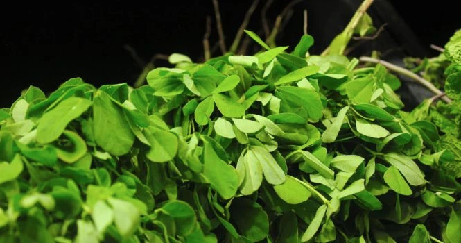
- Coconut water
- Pineapple
- Barley
- Banana
- Lemon
- Horse Gram
- Carrots
- Bitter Gourd
- Increase fluid intake
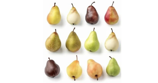
Avoid the following, which are known to increase the formation of stones
- Spinach
- Tomato Seeds
- Black grapes
- Cashew nuts
- Cucumber
- Amla
- Chikoo
- Pumpkin
- Mushroom
- Red Meat
- Fish
- Milk Products
- Brinjal
- Cauliflower
- Gingelly seeds
Frequently Asked Questions
Can kidney stones recur ?
Yes, kidney stones tend to recur . As recurrence is frequent, it’s important to drink 3L of water per day.
Sources
https://www.ncbi.nlm.nih.gov/pmc/articles/PMC6352122/figure/foods-08-00037-f001/?report=objectonly
image : BruceBlaus, CC BY-SA 4.0 https://creativecommons.org/licenses/by-sa/4.0, via Wikimedia Commons
https://www.ncbi.nlm.nih.gov/pmc/articles/PMC3329132/
Image by azwer from Pixabay
Blausen.com staff (2014). “Medical gallery of Blausen Medical 2014”. WikiJournal of Medicine 1 (2). DOI:10.15347/wjm/2014.010. ISSN 2002-4436.De la traducción Ortisa, CC BY-SA 3.0 https://creativecommons.org/licenses/by-sa/3.0, via Wikimedia Commons
Sunshineconnelly at English Wikibooks., CC BY 3.0 Commonshttps://creativecommons.org/licenses/by/3.0, via Wikimedia Commons
© Nevit Dilmen, CC BY-SA 3.0 https://creativecommons.org/licenses/by-sa/3.0, via Wikimedia Commons
![]()
TAGS
Related Articles


Suggested Articles