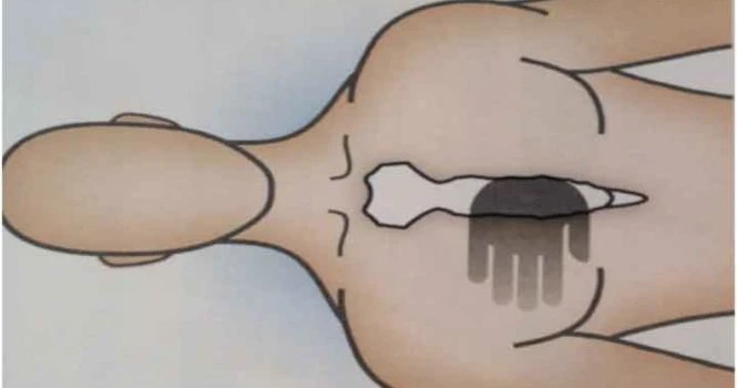
The Human Heart is situated in the center of the chest, behind the sternum.
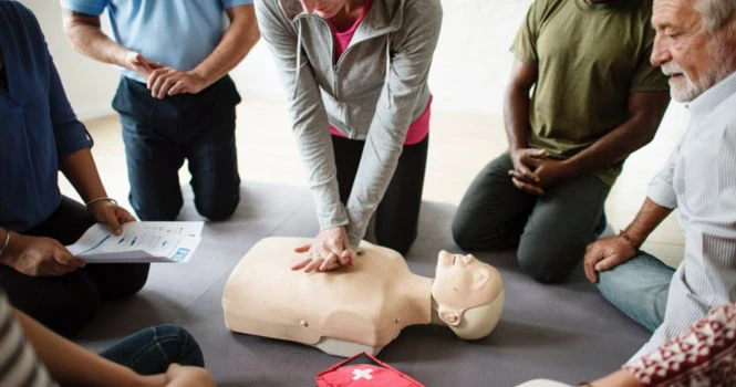
Because of its location, during CPR (Cardio-Pulmonary Resuscitation),we are taught to Push Hard and Push Fast at the lower half of the chest bone (Sternum), to mimic the pumping action of the heart.
Position of Hand during CPR
Chambers of the Heart

It has 4 chambers: 2 atria and 2 ventricles.
Right side of the heart:
Right side of the heart has 2 chambers, the right atrium and the right ventricle.
Right atrium has SA Node (Sinoatrial Node), which is the natural pacemaker of the heart and responsible for triggering impulses, which leads to contraction of the cardiac muscle.
Right atrium receives venous blood, also called deoxygenated blood from Superior Vena cava and Inferior Vena cava.
Right atrium connects with the right ventricle through the Tricuspid valve. Thin walled and low pressure chamber.
Right ventricle is thick walled, and the pulmonary artery arises from the right ventricle.
Left Side of the Heart:
Left side of the heart has 2 chambers, the left atrium and the left ventricle.
Left atrium receives oxygenated blood from the lungs through pulmonary veins.
This is the only exception in the body that a vein (Pulmonary Vein) is carrying oxygenated blood and an artery (Pulmonary artery) is carrying deoxygenated blood.
Left Ventricle is thick walled. Aorta arises from the left ventricle.
Blood Circulation in the Chambers
Blood from all over the body is brought by large veins, namely, superior vena cava and inferior vena cava, which drain into the right atrium; from the right atrium it passes into the right ventricle through the tricuspid valves.
From right ventricle, the blood moves to the lungs through pulmonary artery to get oxygen from the lungs and back into left atrium through pulmonary veins;
From the left atrium, it moves into the left ventricle through bi-cuspid valve.
From the Left ventricle, it pumps all over the body through the aorta and its branches.
It beats non-stop continuously with a regular rhythm. The normal heart rate is 60 beats per minute to 100 beats per minute.
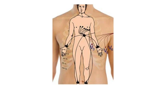
Valves of the Heart
With the atria and major vessels removed, all four valves are clearly visible.

Linings or Layers of wall of the Heart
Heart is made up of 3 layers of tissue,
1. Pericardium (Outer layer)
– Parietal layer of serous pericardium
– Pericardial cavity
– Epicardium (Visceral layer of serous pericardium)
2. Myocardium (Middle muscle layer)
3. Endocardium (Innermost layer)
Pericardium:
It is basically the covering of the heart. It is made up of 2 layers
i) Outer Parietal pericardium
ii) Inner Visceral pericardium
The space between outer parietal pericardium and inner visceral pericardium is called the pericardial space or pericardial cavity and this cavity contains a thin film of fluid.
Outer Parietal Pericardium:
It is a strong protective layer and helps to anchor the heart in the mediastinum.
Parietal Pericardium is made up of 2 layers, the outer fibrous layer and inner serous layer.
Inner Visceral pericardium:
It lines the surface of myocardium and it’s also called Epicardium.
Myocardium
Myocardium is the middle layer of the wall of the heart and is thickest of all and composed of specialized myocardial cells called Myocytes, which contract like other muscles but also conduct electrical impulses to coordinate contraction.
Myocardium has 3 types of muscle fibres:
i) Muscle fibres which form the contractile unit of the heart
ii) Muscle fibres which form the pacemaker
iii) Muscle fibres which form the conductive system of the heart.
Muscle fibres which form the contractile unit of the heart:
Cardiac muscle fibre is branched whereas it’s non-branched in skeletal muscle.
Muscle fibres which form the pacemaker:
Pacemaker is the structure in the heart which generates impulses for heart beat.
Muscle fibres which form the conductive system of the heart:
It is made up of specialized cardiac tissue. Impulses from the atria to the ventricle move through these tissues.
Endocardium:
Endocardium is the innermost lining composed of endothelial cells, which provide a smooth, glistening, non-sticky surface for the smooth flow of blood. Endothelium continues as the endothelium of the blood vessels.
Inflammation of endocardium is called endocarditis.