What is ECG or EKG or Electrocardiogram or Electrocardiograph?
ECG full form is Electrocardiogram or Electrocardiograph. In some countries, the abbreviation EKG is also used.
Electrocardiogram is a screening test, a tool to support a diagnosis.
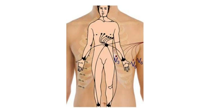
To capture the signals generated by the heart on paper, metal electrodes are placed on the chest surface and extremities and then amplified and recorded by an ECG machine. The signals are compared to the series of specialised algorithms to detect normal from abnormal.
The six limb leads (I, II ,III, VR, VL and VF ) look at the heart from the sides and the feet in a vertical plane.
Leads V1, V2, V3, and V4 as a group effectively view the anterior portion of the heart and are called the an
Uses of ECG:
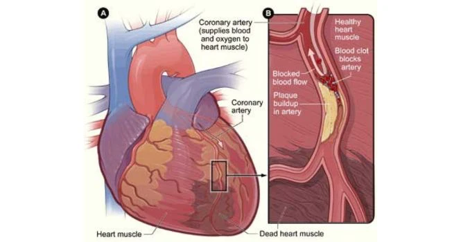
- Its non-invasive, inexpensive and highly useful tool to detect arrhythmias, conduction disturbances and myocardial ischemia (Heart Attack)
- It’s also important to detect other life threatening metabolic disturbances like hyperkalemia (Increased potassium in blood)
- It helps in the diagnosis of the cause of dizziness, syncope and breathlessness.
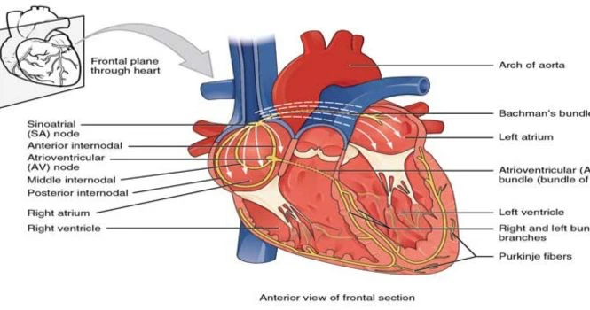
The Electricity of the Heart:
When any muscle contracts, there is depolarisation. If we want to measure the heart’s electrical activity, then the person has to be relaxed as we dont want the skeletal muscle contraction to come in the way. Hence, the person has to be relaxed while taking an ECG.
The Rhythm of the Heart:
The normal heart rhythm is controlled by the SA Node and when the electrical activity starts in the SA Node, then we call it the Sinus Rhythm.
Different Parts of ECG:

There are P, Q, R , S , T and U waves . These names were given arbitrarily and we have been following it .
Time and Speed:
ECG machines record the changes in electrical activity by drawing a trace on a piece of moving paper.
ECG machines run at a standard rate of 25mm/s and use paper with standard-sized squares.
Each large square (5 mm) represents 0.2 second (s), i.e. 200 milliseconds (ms)
Therefore, there are five large squares per second, and 300 per minute.
So an ECG event, such as a QRS complex, occurring once per large square is occurring at a rate of 300/min.
Conduction Problems of the Heart:
- First degree Heart Block
- Second Degree Heart Block
- Third Degree Heart Block
There might be conduction blocks in the right or left bundle branch, called Right Bundle branch block and Left Bundle Branch block (LBBB)
RBBB or Right bundle branch block can be found in normal patients too.
When your Physician or Cardiologist tells you that your ECG is Normal, it means it’s normal at that point in time.
So, patients are usually kept under observation for 24-48 hours, when they report chest discomfort or symptoms relating to coronary disease and also asked to undergo a few blood tests to detect cardiac specific enzymes released in blood by the heart.
Most of the heart conditions are detected in routine health screening camps by ECG.
How is an ECG performed on a Woman
An electrocardiogram (ECG or EKG) is a test that measures the electrical activity of the heart. The procedure is the same for both men and women, but there are a few specific considerations when performing an ECG on a woman.
Here is a general outline of the process:
1. Preparation:
- The patient will be asked to change into a gown or remove clothing from the upper body. For women, it is important to wear a loose-fitting bra or a sports bra that can be easily adjusted or removed.
- The patient will be asked to lie down on an examination table, and the healthcare provider will ensure they are comfortable and relaxed. Any body movement including shivering will give invalid waveforms and it would be repeated again.
2. Electrode placement:
- The technician will clean and, if necessary, shave small areas on the patient’s chest, arms, and legs to ensure proper adhesion of the electrodes.
- For women, special attention will be given to placing the electrodes properly to avoid discomfort and maintain privacy. This may involve placing the electrodes below the breast or, in some cases, lifting the breast to place the electrode underneath.
- A total of 10 electrodes will be attached: six on the chest and one on each limb. The electrodes are connected to the ECG machine via wires.
3. Performing the ECG:
- Once the electrodes are in place, the patient will be asked to lie still and breathe normally while the ECG machine records the electrical activity of the heart.
- The test typically takes only a few minutes and is painless. The ECG machine generates a graph that represents the heart’s electrical activity, which can be printed out or viewed on a screen.
4. After the test:
- The technician will remove the electrodes and clean the areas where they were placed.
- The patient can put their clothes back on and resume normal activities.
The results of the ECG will be reviewed by your Doctor, who will discuss the findings and any further steps to be taken or not.
What information does ECG give about a person?
Some of the key information that an ECG can provide includes:
1. Heart rate: The ECG shows the number of times the heart beats per minute. A normal resting heart rate for adults ranges from 60 to 100 beats per minute.
2. Heart rhythm: The ECG can reveal if the heart is beating regularly or irregularly, helping to identify arrhythmias (abnormal heart rhythms).
3. Cardiac conduction: The ECG can show how electrical impulses travel through the heart, providing information about the conduction system and its function.
4. Heart muscle damage: An ECG can show evidence of past heart attacks or myocardial infarctions by revealing areas of the heart muscle that have been damaged, or heart attack at that moment in time, when the patient comes to ER with chest pain or symptoms of Heart Attack.
5. Ischemia: The ECG can detect areas of the heart muscle that may not be receiving enough oxygen, which could indicate ischemia or a potential risk of a heart attack.
6. Chamber enlargement: An ECG can reveal if the heart’s chambers (atria or ventricles) are enlarged, which may suggest conditions like cardiomyopathy, heart failure, or valve disease.
7. Electrolyte imbalances: Abnormal ECG findings can sometimes indicate electrolyte imbalances (e.g., potassium or calcium levels), which can affect the heart’s function.
8. Medication effects: The ECG can show the effects of certain medications on the heart, helping healthcare providers monitor their patients’ response to treatment.
While an ECG can provide essential information about a person’s heart health, it is not a comprehensive diagnostic tool. It is a Screening Test.
Additional tests or evaluations may be required to confirm a diagnosis or to gather more information about a particular condition.