Neuron, also called the nerve cell, is the functional unit of nervous system.
Abundant during birth and shortly thereafter, they tend to remain constant throughout life and once they die they don’t regenerate nor divide unlike other cells of the body.
Classification of Neurons:
Neurons are classified by three different methods,
1. Based on number of poles
2. Based upon function
3. Based upon the length of axon
1. Based on number of poles:
On the basis of number of poles they possess,they are divided into three types
– Unipolar Neurons
– Bipolar Neurons
– Multipolar Neurons
Unipolar Neurons:
Uni means one . These neurons have only one pole. Axon and Dendrites arise from a single pole. THese type of neurons are present only in the embryonic stage in humans.
Bipolar Neurons:
Bi means two. These neurons have 2 poles. Axons arise from one pole, while the dendrites arise from other pole.
Multipolar Neurons:
Multi means Many. These type of neurons have many poles. While one of the pole gives rise to Axon, and all other poles give rise to dendrites.
2. Based upon function:
On the basis of function they are divided into two types
– Motor or Efferent neurons
– Sensory or afferent neurons
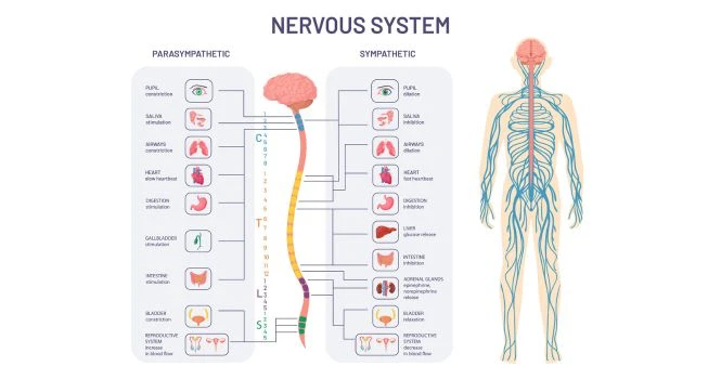
Motor or Efferent neurons
Motor neurons send motor commands from the cortex to the spinal cord or from the spinal cord to the muscles; or in other words from the central nervous system to the peripheral effector organs like the muscles, glands, blood vessels etc.
Generally, each motor neuron has a long axon and short dendrite.
Sensory or afferent neurons
Sensory neurons, receive signals from all sensory organs and transmit them to central nervous system through the short axons ;
Generally each sensory neuron has a short axon and long dendrites.
3. Based upon the length of axon
– Golgi type I neuron
– Golgi type II neuron
Golgi Type I Neuron:
These types of neurons have long Axons. While the cell body is in different parts of the central nervous system, while their axons reach the remote peripheral organs.
Golgi Type II Neuron:
These types of neurons have short Axons. They are present in Brain ( cerebral cortex ) and spinal cord.
STRUCTURE OF NEURON
Neuron is made up of
- Nerve Cell Body
- Dendrite
- Axon
Dendrites and Axons are processes of neuron. Dendrites are short processes while axons are long processes.
Dendrites and Axons are commonly called Nerve Fibres.
NERVE CELL BODY
Synonyms of Nerve Cell Body are Soma or Perikaryon
It’s irregular in shape and has a cytoplasm called neuroplasm, covered with a cell membrane.
The cytoplasm has large nucleus, Nissl Bodies, neurofibrils, mitochondria and Golgi apparatus.
Nissl bodies and neurofibrils are found only in nerve cells and not in any other cell of the body.
Nucleus:
Each neuron has one centrally placed Nucleus, with one or more prominent nucleoli.
The nerve cell cannot multiply like other cells because it doesn’t have a centrosome.
Centrosome is the structure involved in cell division.
Nissl Bodies:
Nissl bodies or Nissl granules are found in cytoplasm of Neuron and are present in soma and dendrite, but not in axon or axon hillock.
Nissl granules are called tigroid substances because of the spotted appearance of soma ( Tigroid means spotted or striped appearance ), after suitable staining.
Nissl Granules are found in Dendrites. They also have ribosomes for protein synthesis.
Number of Nissl bodies in a nerve varies according to the condition of the nerve. During fatigue or injury to the neuron, these bodies fragment and disappear by a process called chromatolysis. Granules appear after the recovery of fatigue or regeneration of nerve fibres.
Neurofibrils:
Neurofibrils are thread-like features characteristic of neuron. They consist of microfilaments and microtubules.
Mitochondria:
These powerhouse of the cell are present in soma and axon.
Golgi Apparatus:
This is involved in packaging and processing of proteins into granules.
DENDRITE
Dendrite is a branched process of neurons and it is branched repeatedly.
They are tree-like projections,which receive information from other neurons.They make connections with other neurons through synapses.
- Most neurons have many dendrites
- However,some neurons may have only one dendrite
- Short and highly branched
- Transmits information to the cell body.
The terminal buttons are located at the end of the neuron and are responsible for sending the signal on to other neurons. At the end of the terminal button is a gap, known as synapse.
Synapses act as a relay station transmitting signals from one neuron to another.
AXON
Axon is a longer process of Nerve cells.
They are long, thin projections from the cell body of neuron..
- Most neurons have only one axon
- Information is transmitted away from the cell body.
- Myelin covering may or may not be present.
- Myelin acts as an insulator for some axons.
Organisation of Nerve:
Each nerve is formed by many bundles of nerve fibres and each bundle of nerve fibres is called a Fasciculus.
Covering of Nerve:
Each nerve Fibre is covered with Endoneurium.
Nerve fibre Bundle is called Fasciculus.
Fasciculus is covered with Perineurium.
The whole nerve is covered by Epineurium.
Internal structure of Axon- Axis Cylinder:
Axon has a long central core of cytoplasm called axoplasm.
Axoplasm is covered with axolemma. It is the continuation of the cell membrane of the cell body.
Axoplasms along with axolemma are called the Axis cylinder of the nerve fibre.
Axis cylinder of the nerve fibre is covered by a membrane called neurilemma.
Non-myelinated Nerve Fibre:
These Nerve fibres are not covered by Myelin sheath.
Myelinated Nerve Fibre:
Nerve fibers insulated by Myelin sheath are Myelinated Nerve Fibre.
MYELIN SHEATH
Myelin sheath is responsible for the white color of the nerve fibre.
It is a thick lipoprotein sheath, which insulates the myelinated nerve fibre. It is not continuous and absent at regular intervals. The area where myelin sheath is absent is called Nodes of Ranvier. Segment of nerve fibre between two nodes is called internode.
Chemistry of Myelin Sheath:
Myelin sheath is formed by concentric layers of proteins layed with Lipids.
Lipids are Cholesterol, lecithin and cerebroside (Sphingomyelin).
Formation of Myelin Sheath – Myelinogenesis
Formation of myelin sheath around the axon is called myelinogenesis.It is formed by schwann cells in neurilemma . Myelinogenesis in the peripheral nerves , starts in the 4 month of intrauterine life and is completed only in the second year of life.
Functions of Myelin Sheath
- Faster conduction
In myelinated nerve fibres, the impulses jump from one node to another node. This type of transmission of impulses is called Saltatory conduction.
- Insulating Capacity
Because of it’s high insulating capacity, it restricts the nerve impulses from stimulating the neighbouring nerve fibres.
NEURILEMMA:
Also called Neurilemmal Sheath or Sheath of Schwann.
It’s a thin membrane which surrounds the Axis cylinder.
Functions of Neurilemma:
Neurilemma is absent in the central nervous system, so, neuroglial cells called oligodendroglia are responsible for myelinogenesis in the central nervous system.
- In Non-myelinated nerve fibre, neurilemma serves as a covering membrane.
- In myelinated nerve fibre, it is necessary for the formation of myelin sheath.
Questions and Answers
What do dendrites do in a neuron?
Dendrites are the branched projections of a neuron that receive information from other neurons or sensory receptors. Their main function is to transmit electrical signals and chemical messages (neurotransmitters) from other neurons to the cell body of the neuron, where the signals are integrated and processed.
Dendrites play a crucial role in the communication between neurons, as they receive incoming signals from other neurons and transfer them to the cell body. They are covered in synapses, which are the connections between neurons that allow for the transmission of electrical or chemical signals. The more synapses a neuron has on its dendrites, the more information it can receive and process.
Dendrites also have specialized structures called dendritic spines, which are small protrusions that increase the surface area of the dendrites and provide more space for synapses to form. The number and shape of dendritic spines can change in response to experience, learning, and memory formation, which is thought to play a role in neural plasticity.
Overall, dendrites play a crucial role in the communication and processing of information within the nervous system, allowing for complex signaling and neural plasticity.