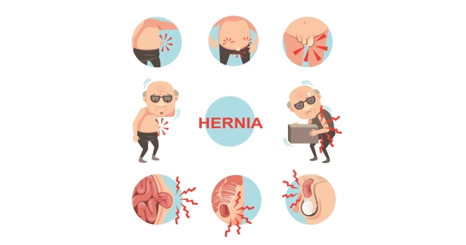
“The Patient groans in agony”, is the phrase often associated with pain in Kidney Stones.
Let’s see how to deal with them and the options we have.
Kidney stones are small, hard mineral deposits that form inside your kidneys, causing a myriad of symptoms ranging from mild discomfort to severe pain. While these tiny stones can lead to significant health issues if not properly addressed.
Definition of Kidney Stones or Renal Stones
Kidney stones are solid masses made up of crystals that originate within the kidneys.
These stones are composed of various substances, the most common being calcium oxalate. Other types include struvite, uric acid, and cystine stones. They can vary in size, from as small as a grain of sand to as large as a golf ball.
Kidney stones form when the balance of water, minerals, and salts in the urine changes, leading to the crystallization of these substances around a small nucleus, which could be a tiny piece of calcified tissue or a microscopic crystal.
The presence of kidney stones can lead to symptoms such as severe pain in the abdomen or flank, blood in the urine, frequent urination, and nausea or vomiting.
However, some stones may be asymptomatic and only discovered during routine medical check-ups. Treatment can range from simple hydration and pain management to surgical procedures for larger, more problematic stones.
Understanding the types, causes, and symptoms of kidney stones is crucial for effective prevention and treatment, which is why a comprehensive guide can be an invaluable resource for patients.
Causes of Kidney Stones
Understanding the root causes of kidney stones is crucial for both prevention and treatment. While the formation of kidney stones can be influenced by a variety of factors, some of the most common causes include:
A. Mineral Build-up
Mineral build-up occurs when substances like calcium, oxalate, and phosphorus become concentrated in the urine. This can lead to the crystallization of these substances, forming the nucleus around which a kidney stone may grow.
Contributing Factors:
- Dehydration: Lack of adequate fluid intake can concentrate minerals in the urine.
- High mineral content in drinking water: Some geographical areas have water that is rich in minerals, contributing to stone formation.
B. Dietary Factors
Certain foods and drinks can contribute to the formation of kidney stones by increasing the levels of stone-forming substances in the urine.
Contributing Factors:
- High sodium intake: Excessive salt can increase calcium levels in the urine.
- Oxalate-rich foods: Foods like spinach, rhubarb, and beets are high in oxalates.
- Animal protein: Diets high in animal protein can increase uric acid and reduce citrate in the urine, both of which can contribute to stone formation.
- Low fluid intake: Not drinking enough water can concentrate the urine, making stone formation more likely.
C. Genetic Factors
Some individuals are genetically predisposed to kidney stone formation. A family history of kidney stones can significantly increase one’s risk.
Contributing Factors:
- Inherited metabolic disorders: Conditions like cystinuria and primary hyperoxaluria are genetic disorders that can lead to kidney stones.
- Family history: A family history of kidney stones can be a strong indicator of one’s risk.
D. Medical Conditions
Certain medical conditions can make an individual more susceptible to developing kidney stones.
Contributing Factors:
- Hyperparathyroidism: Overactivity of the parathyroid glands can lead to increased calcium levels in the urine.
- Urinary tract infections: Infections can lead to the formation of struvite stones.
- Inflammatory bowel disease: Conditions like Crohn’s disease can affect the absorption of calcium and water, contributing to stone formation.
- Renal tubular acidosis: This condition alters the acid-base balance in the body, leading to the formation of stones.
Types of Kidney Stones
1. Calcium oxalate Stones
Composition:
Calcium oxalate stones are the most common type of kidney stone, accounting for about 80% of all cases. These stones are primarily composed of calcium oxalate, a naturally occurring compound found in certain foods and also produced by the liver.
Causes:
Factors contributing to the formation of calcium oxalate stones include:
- High levels of calcium, oxalate, or both in the urine
- Dehydration, which concentrates the urine
- High intake of oxalate-rich foods like spinach, rhubarb, and beets
- Certain metabolic disorders, such as primary hyperoxaluria
- Overuse of Vitamin D supplements
Symptoms:
The symptoms are similar to those of other kidney stones and may include:
- Severe pain in the back or sides
- Blood in the urine (hematuria)
- Frequent urination
- Nausea and vomiting
Diagnosis:
Calcium oxalate stones are usually diagnosed through imaging tests like ultrasound, X-ray, or CT scans. Urinalysis and blood tests may also be conducted to determine the levels of calcium and oxalate in the body.
Treatment:
Treatment options include:
- Increased fluid intake to help pass the stone
- Medications to relax the ureter and facilitate stone passage
- Lithotripsy, a procedure that uses shock waves to break up the stone
- Surgical removal for larger stones
Prevention:
Preventive measures may include:
- Drinking plenty of water to dilute urine
- Reducing intake of oxalate-rich foods
- Calcium supplements, but only under medical supervision
- Medications like thiazide diuretics to reduce calcium levels in urine
2. Calcium Phosphate Stones
Composition:
Calcium phosphate stones are another type of calcium-based kidney stone, although they are less common than calcium oxalate stones. These stones are primarily composed of calcium and phosphate ions.
Causes:
Factors contributing to the formation of calcium phosphate stones include:
- Alkaline Urine: A higher pH level in the urine can promote the formation of calcium phosphate stones.
- Hyperparathyroidism: Overactivity of the parathyroid glands can lead to elevated levels of calcium in the urine.
- Renal Tubular Acidosis: This condition can lead to an alkaline urine environment, which is conducive for calcium phosphate stone formation.
Symptoms:
The symptoms of calcium phosphate stones are similar to those of other types of kidney stones, such as:
- Severe pain in the back, abdomen, or groin
- Blood in the urine (hematuria)
- Frequent urination
- Nausea and vomiting
Diagnosis:
Calcium phosphate stones can be diagnosed through various imaging tests like X-rays, ultrasounds, or CT scans. Urinalysis and blood tests may also be used to check for elevated levels of calcium and phosphate.
Treatment:
Treatment options for calcium phosphate stones are similar to those for other types of kidney stones:
- Increased fluid intake: To dilute the concentration of minerals in the urine.
- Medications: Such as thiazide diuretics to reduce calcium levels in the urine.
- Lithotripsy: A non-invasive procedure that uses shock waves to break up the stone.
- Surgical removal: For larger or more problematic stones.
Prevention:
Preventive measures for calcium phosphate stones may include:
- Maintaining a balanced diet: Low in sodium and high in fluids.
- Medications: Such as potassium citrate to regulate urine pH.
- Monitoring: Regular check-ups and urine tests to monitor calcium and phosphate levels.
3. Struvite ( Magnesium ammonium Phosphate) Stones
Composition:
Struvite stones are composed of magnesium, ammonium, and phosphate. They are also commonly known as “infection stones” because they often form in conjunction with urinary tract infections (UTIs).
Causes:
The primary factors contributing to the formation of struvite stones include:
- Urinary Tract Infections: Struvite stones often form as a result of UTIs caused by bacteria that produce an enzyme called urease. This enzyme breaks down urea in the urine into ammonia, making the urine more alkaline and conducive to struvite stone formation.
- Alkaline Urine: A higher pH level in the urine can promote the crystallization of magnesium, ammonium, and phosphate, leading to struvite stones.
Symptoms:
The symptoms of struvite stones can be similar to other types of kidney stones but may also include symptoms of a UTI, such as:
- Pain or burning during urination
- Cloudy or foul-smelling urine
- Fever and chills
- Severe pain in the back, abdomen, or groin
- Frequent urination
Diagnosis:
Struvite stones are usually diagnosed through imaging tests like ultrasounds or CT scans. A urinalysis may also be conducted to check for signs of infection and the presence of struvite crystals.
Treatment:
Because struvite stones are often associated with UTIs, treatment usually involves:
- Antibiotics: To treat the underlying infection.
- Surgical Removal: Struvite stones can grow quite large and may require surgical intervention.
- Lithotripsy: For smaller stones, shock wave lithotripsy may be used to break up the stones into smaller pieces that can be passed more easily.
Prevention:
Preventive measures for struvite stones focus on:
- UTI Prevention: Regular urination, proper hygiene, and in some cases, prophylactic antibiotics can help prevent UTIs.
- Acidifying Urine: Certain medications can help make the urine more acidic, which can prevent the formation of struvite stones.
- Regular Monitoring: Frequent check-ups and urine tests can help monitor for signs of UTIs and stone formation.
4. Uric Acid Stones
Composition:
Uric acid stones are formed from uric acid, a byproduct of the breakdown of purines—substances found in certain foods and naturally occurring in the body. Unlike other types of kidney stones, uric acid stones are more soluble in alkaline environments and less soluble in acidic conditions.
Causes:
Factors contributing to the formation of uric acid stones include:
- Acidic Urine: A lower pH level in the urine can promote the crystallization of uric acid.
- High-Purine Diet: Consuming foods rich in purines like red meat, shellfish, and some types of fish can increase uric acid levels.
- Gout: This condition, characterized by high levels of uric acid in the blood, can also lead to uric acid stones.
- Dehydration: Concentrated urine can increase the risk of all types of kidney stones, including uric acid stones.
Symptoms:
The symptoms of uric acid stones are similar to those of other kidney stones:
- Severe pain in the back, abdomen, or groin
- Blood in the urine (hematuria)
- Frequent urination
- Nausea and vomiting
Diagnosis:
Uric acid stones may not be easily visible on standard X-rays, making ultrasounds and CT scans more effective for diagnosis. Urinalysis and blood tests can also help identify elevated levels of uric acid.
Treatment:
Treatment options for uric acid stones include:
- Alkalinization of Urine: Medications like potassium citrate can make the urine more alkaline, helping to dissolve uric acid stones.
- Increased Fluid Intake: To dilute the concentration of uric acid in the urine.
- Dietary Changes: A low-purine diet can help reduce uric acid levels.
- Medications: Drugs like allopurinol can be used to control uric acid levels in people with gout or other conditions.
Prevention:
Preventive measures for uric acid stones may include:
- Drinking plenty of water to dilute the urine
- Avoiding high-purine foods
- Regular monitoring of uric acid levels through blood and urine tests
- Medications to control uric acid levels, if necessary
5. Cystine Stones
Composition:
Cystine stones are formed from cystine, a type of amino acid that is a building block of proteins. These stones are relatively rare and usually occur in individuals with a genetic disorder known as cystinuria, which affects the reabsorption of cystine in the kidneys.
Causes:
The primary factor contributing to the formation of cystine stones is:
- Cystinuria: This inherited metabolic disorder leads to high levels of cystine in the urine, which can crystallize and form stones.
Symptoms:
The symptoms of cystine stones are similar to those of other types of kidney stones:
- Severe pain in the back, abdomen, or groin
- Blood in the urine (hematuria)
- Frequent urination
- Nausea and vomiting
Diagnosis:
Cystine stones are usually diagnosed through specialized tests, including:
- Urinalysis: To check for high levels of cystine.
- 24-hour urine collection: To measure the total amount of cystine.
- Imaging Tests: Like ultrasounds or CT scans to locate the stones.
Treatment:
Treatment options for cystine stones are more specialized and may include:
- Increased Fluid Intake: To dilute the concentration of cystine in the urine.
- Alkalinization of Urine: Medications like potassium citrate can make the urine more alkaline, which may help to dissolve cystine stones.
- Medications: Drugs that bind to cystine and make it more soluble may be used.
- Surgical Removal: For larger or more problematic stones, surgical intervention may be necessary.
Prevention:
Preventive measures for cystine stones focus on:
- High Fluid Intake: To keep the urine diluted.
- Regular Monitoring: Frequent check-ups and urine tests to monitor cystine levels.
- Dietary Changes: A balanced diet low in sodium may help reduce cystine stone formation.
- Medications: Such as cystine-binding thiol drugs can be used to make cystine more soluble in urine.
Diagnosis of Kidney Stones
Diagnostic Procedures
1. Imaging Tests
Imaging tests are among the most commonly used methods to detect, locate, and characterize kidney stones. They can provide detailed images of the urinary tract and help determine the size, shape, and number of stones present.
A. X-rays (KUB – Kidneys, Ureters, Bladder):
- Purpose: Traditional X-rays can detect most kidney stones, especially those containing calcium.
- Limitations: Uric acid stones and some other types may not be visible on standard X-rays.
B. Ultrasound:
- This is the first investigation carried out in Kidney Stones
- Purpose: This test uses sound waves to produce images and can detect kidney stones in both the kidneys and ureters. It’s a preferred method during pregnancy due to the absence of radiation.
- Limitations: Might not detect very small stones as clearly as other imaging methods.
C. CT Scan (Computed Tomography):
- This is the investigation of choice in Kidney Stones
- Purpose: Non-contrast helical CT scans are particularly useful in detecting stones, providing detailed cross-sectional images of the body. They can detect almost all types of kidney stones and are considered one of the most reliable methods for diagnosing kidney stones.
- Limitations: Involves radiation exposure, which may not be suitable for everyone, especially pregnant women.
D. Intravenous Pyelogram (IVP) or Intravenous Urogram (IVU):
- Purpose: A dye is injected into a vein, and X-rays are taken as the dye travels through the kidneys, ureters, and bladder. This test can show the size, shape, and position of kidney stones and any obstructions.
- Limitations: Involves exposure to both dye (which can cause allergic reactions in some people) and radiation. It’s less commonly used today due to the effectiveness and convenience of CT scans.
E. Magnetic Resonance Imaging (MRI):
- Purpose: While not a standard method for diagnosing kidney stones, MRI can be used in special cases to visualize renal anatomy and stones.
- Limitations: More expensive and not as widely available for this specific purpose. Might not offer any significant advantage over CT scans for most stones.
In addition to imaging tests, a doctor may also order blood tests to determine the levels of certain minerals that can contribute to stone formation, and urine tests to check for the presence of crystals and to determine urine pH.
The choice of diagnostic procedure often depends on the patient’s specific situation, the suspected type of stone, and the resources available.
2. Blood and Urine Tests
Blood and urine tests are often used in conjunction with imaging studies to provide a comprehensive diagnosis of kidney stones. These tests can offer insights into the underlying causes of stone formation and guide treatment and prevention strategies.
A. Blood Tests:
- Purpose: Blood tests can measure levels of certain minerals like calcium, phosphate, and uric acid, as well as assess kidney function. Elevated levels of these substances may indicate an increased risk for stone formation.
- Common Tests:
- Complete Blood Count (CBC)
- Blood Urea Nitrogen (BUN)
- Creatinine
- Calcium
- Phosphate
- Uric Acid
- Limitations: While blood tests can indicate an increased risk for stone formation, they cannot confirm the presence of a stone.
B. Urine Tests:
- Purpose: Urine tests can identify crystals, red and white blood cells, and bacteria in the urine. They can also measure urine pH, which can indicate the type of stone you’re most likely to form.
- Common Tests:
- Urinalysis: A quick test that can detect blood, crystals, and other substances in the urine.
- 24-Hour Urine Collection: This test measures the amount of various substances in urine, including those that can form stones, over a 24-hour period.
- Limitations: Urine tests are generally more useful for determining the cause of kidney stones and guiding treatment rather than for acute diagnosis.
C. Urine Culture:
- Purpose: If a urinary tract infection (UTI) is suspected—common with struvite stones—a urine culture can identify the type of bacteria present to guide antibiotic treatment.
- Limitations: A urine culture is specific to diagnosing infections and won’t confirm the presence of a stone.
Treatment Options for Kidney Stones
A. Medical Treatments
Medical treatments for kidney stones aim to relieve symptoms, facilitate the passage of the stone, and prevent future stone formation. Here are some of the most commonly used medical treatments:
1. Medications
A. Pain Relievers:
- Purpose: To manage the often severe pain associated with kidney stones.
- Common Options: Over-the-counter pain relievers like ibuprofen, or prescription medications such as opioids for more severe pain.
B. Alpha Blockers:
- Purpose: To relax the muscles in the ureter, helping the stone to pass more easily.
- Common Options: Tamsulosin (Flomax) is frequently used for this purpose.
C. Antibiotics:
- Purpose: To treat any underlying urinary tract infections, often associated with struvite stones.
- Common Options: Depends on the type of bacteria found in urine culture.
D. Medications to Control Stone Formation:
- Purpose: To prevent future stone formation by controlling levels of certain substances in the urine.
- Common Options: Thiazide diuretics to reduce calcium in urine, allopurinol for uric acid stones, and potassium citrate to alkalize the urine.
2. Lithotripsy
A. Extracorporeal Shock Wave Lithotripsy (ESWL):
- Purpose: To break up kidney stones into smaller pieces using shock waves, making them easier to pass.
- Procedure: The patient lies on a water-filled cushion, and shock waves are directed at the stones from outside the body.
- Limitations: Not effective for very large stones or for stones located in certain positions. May require multiple sessions.
B. Laser Lithotripsy:
- Purpose: To break up stones using laser energy.
- Procedure: A ureteroscope is inserted into the ureter, and laser energy is directed at the stone to break it into smaller pieces.
- Limitations: Invasive procedure that may require anesthesia and has a risk of complications like infection or ureteral injury.
C. Electrohydraulic Lithotripsy (EHL):
- Purpose: To break up stones using electrical shocks.
- Procedure: Similar to laser lithotripsy but uses electrical energy instead of laser energy.
- Limitations: Less commonly used than other forms of lithotripsy and may require anesthesia.
Each treatment option has its own set of advantages and limitations, and the best treatment for you will depend on the type, size, and location of your kidney stones, as well as your overall health.
Always consult with your Doctor for a diagnosis and treatment plan tailored to your individual needs.
B Surgical Options
When kidney stones are too large to pass naturally, cause severe pain, or lead to complications like urinary tract infections, surgical intervention may be necessary. Here are some common surgical options for treating kidney stones:
1. Ureteroscopy
- Purpose: To remove stones located in the ureter or kidney.
- Procedure: A thin, flexible scope (ureteroscope) is inserted through the urethra and bladder into the ureter. Small stones can be removed directly, while larger ones may be broken up using laser or other energy sources before removal.
- Limitations: May not be suitable for very large stones or stones located in certain positions within the kidney.
2. Percutaneous Nephrolithotomy (PCNL)
- Purpose: To remove larger stones or multiple stones in the kidney.
- Procedure: A small incision is made in the back, and a nephroscope is inserted directly into the kidney to remove the stone.
- Limitations: More invasive than other procedures and generally requires hospitalization and anesthesia. There’s a higher risk of complications like bleeding or infection.
3. Laparoscopic Stone Surgery
- Purpose: To remove stones that are not accessible through other methods.
- Procedure: Small incisions are made in the abdomen to insert laparoscopic instruments, which are used to remove the stone.
- Limitations: Highly invasive and generally reserved for complex cases where other methods are not feasible.
4. Open Surgery
- Purpose: To remove extremely large stones or in cases where other treatments have failed.
- Procedure: An incision is made in the back or abdomen to access the kidney and remove the stone.
- Limitations: Most invasive option with a longer recovery time and higher risk of complications. Rarely used nowadays due to the effectiveness of less invasive methods.
5. Robotic-Assisted Surgery
- Purpose: To remove complex or multiple stones with high precision.
- Procedure: Similar to laparoscopic surgery but performed using a robotic surgical system, which allows for more precise movements.
- Limitations: Expensive and not widely available. Generally reserved for complex cases.
Surgical options are generally considered when medical treatments fail or are not suitable due to the size, location, or type of stone.
The choice of surgical method will depend on various factors, including the patient’s overall health and the surgeon’s expertise.
C. Home Remedies and Lifestyle Changes
While medical and surgical treatments are often necessary for treating kidney stones, home remedies and lifestyle changes can play a significant role in both managing current symptoms and preventing future stones. Here are some commonly recommended approaches:
1. Increased Fluid Intake
- Purpose: To dilute the urine, making it less likely for stones to form.
- Recommendation: Aim for at least 2.5 to 3 liters of water per day, unless advised otherwise by a healthcare provider.
2. Dietary Changes
- Purpose: To control levels of substances that contribute to stone formation.
- Recommendation:
- Reduce sodium intake.
- Limit animal protein.
- Avoid oxalate-rich foods if prone to calcium oxalate stones.
- Increase intake of citrate-rich foods like citrus fruits.
3. Exercise
- Purpose: Regular exercise can help prevent kidney stones by maintaining healthy blood pressure levels and efficient kidney function.
- Recommendation: Aim for at least 30 minutes of moderate exercise most days of the week.
4. Herbal Remedies
- Purpose: Some herbs are believed to help prevent kidney stones or aid in their removal.
- Common Options:
- Chanca Piedra: Known as the “stone breaker,” this herb is often used in South America to help dissolve kidney stones.
- Dandelion Root: Believed to help detoxify the kidneys.
- Limitations: The effectiveness of herbal remedies is not universally supported by scientific evidence. Always consult a healthcare provider before starting any herbal treatment.
5. Lemon Juice and Olive Oil
- The citrate in lemon juice can help dissolve calcium-based stones, and olive oil may act as a lubricant for easier passage.
6. Apple Cider Vinegar
- Purpose: Believed to help dissolve kidney stones and alkalize the blood and urine.
- Recommendation: Mix two tablespoons of apple cider vinegar with water and drink daily.
- Limitations: High concentrations of apple cider vinegar can be harmful. Always dilute it and consult a healthcare provider before starting this remedy.
7. Avoiding Excessive Vitamin C and Calcium Supplements
- Excessive supplementation can contribute to stone formation.
- Recommendation: Stick to dietary sources of these nutrients when possible, and consult a healthcare provider if you think you need supplements.
Home remedies and lifestyle changes are generally more effective for prevention than for treating existing stones. Always consult a healthcare provider for a comprehensive treatment plan tailored to your specific condition.
D. Monitoring and Follow-Up Care
After initial treatment for kidney stones, ongoing care is essential to monitor for any recurring stones, complications, or underlying conditions that may contribute to stone formation. Here are some key aspects of monitoring and follow-up care:
1. Regular Check-ups
- To assess kidney function and the potential for recurrent stones.
- Periodic appointments with a healthcare provider for physical examinations, symptom reviews, and discussion of any new risk factors.
2. Repeat Imaging Studies
- To check for the presence of new or remaining stones.
- Common Options: Ultrasound may be repeated at intervals to monitor for stone formation.
3. Blood Tests
- To monitor levels of calcium, uric acid, and other substances that can contribute to stone formation.
- Periodic blood tests to assess kidney function and mineral levels.
4. 24-Hour Urine Collection
- To measure the amounts of various substances in the urine that contribute to stone formation.
- What to Expect: You may be asked to collect your urine over a 24-hour period for detailed analysis.
5. Medication Review
- To adjust any medications that may contribute to stone formation or to introduce new medications that can prevent stones.
6. Lifestyle and Diet Review
- To assess the effectiveness of lifestyle and dietary changes in preventing new stones.
- Ongoing dietary counseling and possibly working with a dietitian to make effective and sustainable changes.
Prevention Strategies for Kidney Stones
Preventing kidney stones often involves a multi-faceted approach that includes dietary modifications, proper hydration, medications, and regular medical check-ups. Here are some key strategies:
A. Dietary Modifications
- Limit Sodium Intake: High sodium levels can increase calcium in your urine. Aim to consume no more than 2,300 mg of sodium per day.
- Reduce Animal Protein: Diets high in animal protein can increase uric acid levels and reduce citrate in the urine, both of which can contribute to stone formation.
- Oxalate-Rich Foods: If you’re prone to calcium oxalate stones, you may need to avoid or limit foods high in oxalates, such as spinach, rhubarb, and beets.
- Increase Citrate Levels: Foods high in citrate, such as citrus fruits, can help prevent stone formation.
- Balanced Calcium Intake: While it may seem counterintuitive, a diet adequate in calcium can actually help prevent calcium oxalate stones.
B. Hydration
- Water Intake: Aim for at least 2.5 to 3 liters of water per day to dilute urine and reduce the concentration of stone-forming minerals.
- Limit Caffeine and Alcohol: Both can contribute to dehydration, increasing the risk of stones.
- Monitor Urine Color: A light, straw-colored urine typically indicates proper hydration.
C. Medications (if applicable)
- Thiazide Diuretics: Used to prevent calcium stones by reducing the amount of calcium released by the kidneys into the urine.
- Potassium Citrate: Helps to alkalize the urine and dissolve certain types of stones.
- Allopurinol: Used for uric acid stones, it helps to lower levels of uric acid in the blood and urine.
- Antibiotics: For struvite stones caused by urinary tract infections, long-term antibiotics may be prescribed to prevent recurrence.
D. Regular Medical Check-ups
- Routine Tests: Regular blood and urine tests can monitor levels of substances that cause stones.
- Imaging Studies: Periodic imaging tests like ultrasounds or X-rays can be useful for those with a history of kidney stones to detect any new stones early.
- Consultations: Regular appointments with healthcare providers for reviews of medical history, medications, and symptoms can help adapt prevention strategies over time.
- Dietary Review: Ongoing consultations with a dietitian can help tailor a diet that minimizes the risk of stone formation.
By implementing these prevention strategies, you can significantly reduce your risk of developing kidney stones. Always consult with a healthcare provider for a prevention plan tailored to your specific needs and conditions.