Conductive system of the heart is by modified cardiac muscle fibres. These are specialised cells which carry impulses from the SA Node to the ventricles.
Components of Conductive system of Human Heart:
- SA Node
- AV Node
- Bundle of HIS
- Right and left bundle branches
- Purkinje fibres
Electrical Activity of the heart:
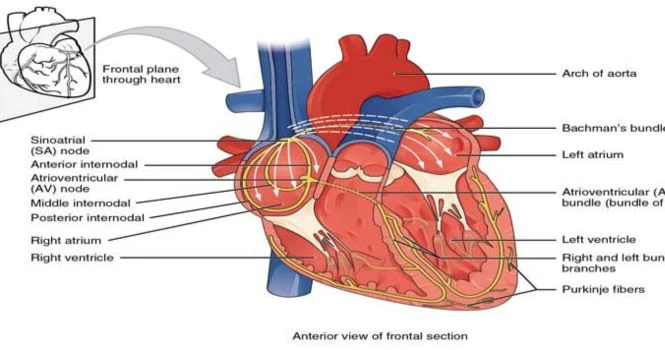
Under normal conditions, the one factor which makes the heart beat non-stop continuously, is the triggering of neural impulses in the SA Node (Sino-atrial node).
SA Node is situated at the junction of the right atrium and the superior vena cava.
Neural impulse from the SA node traverses the right atrium to reach the AV node (Atrio-ventricular node).
AV Node lies at the base of interatrial septum, just above the tricuspid annulus, anterior to the coronary sinus.
The sequence of electrical impulses conducted in the heart is as follows:
SA Node → AV Node → Bundle of HIS → Left bundle branches and right bundle branches→ Purkinje Fibres
SA Node is under the control of inhibitory impulses from the Vagus nerve, which keep the triggering of impulses at a lower level, sufficient for the heart to function optimally and the heart rate to be in normal range. Hypothetically, if we remove the inhibitory impulses from the vagus nerves, then the heart range would be more than 300 beats per minute and fatal.
What is the normal Heart rate in humans?
Normal Heart rate is 60 beats to 100 beats per minute.
If it’s lower than 60, then we call it Bradycardia.
If it’s higher than 100, then we call it Tachycardia.
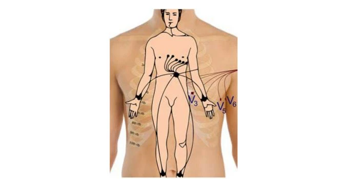
The electrical potentials generated from the specialized cardiac cells can be recorded with electrodes placed on the surface of the chest and connected to a machine called ECG or EKG (Electrocardiogram).
Velocity of impulses at different parts of the conductive system:
- Atrial muscle fibres : 0.3 meters/second
- Internodal fibres : 1.0 meter/second
- AV Node : 0.05 meter/second
- Bundle of HIS : 0.12 meter/second
- Purkinje fibres : 4.0 meter/second
- Ventricular muscle fibres : 0.5 meter/second
The velocity is minimum in AV Node and maximum in Purkinje fibres.
Under resting conditions, myocardial cells are polarized, which means they carry a charge due to transmembrane ion concentration difference.
If the neural impulses cannot pass through the designated pathways in the conduction system, then we call them conduction blocks namely, right bundle branch block (also called RBBB) and left bundle branch block (also called LBBB) and other intermediate and smaller blocks.
Pacemakers are required if the conduction system is not firing impulses for optimal contraction of the heart, so as to yield an effective cardiac output. They can be placed on the surface of the body or internally below the skin.
Contractile Properties of Cardiac muscle:
- Follows ALL or NONE Law
- Staircase Phenomenon
- Summation of Subliminal stimuli
- Refractory Period
ALL OF NONE Law:
Whatever might be the strength of stimulus applied, the whole cardiac muscle gives maximum response, or No response at all.
Below the threshold level, if the strength of stimulus is not adequate then muscle does not give response at all.